MENU
BE | EUR

BE | EUR
-
- All Pipettes, Dispensers & Automated Liquid Handlers
- Mechanical Pipettes
- Electronic Pipettes
- Multi-Channel Pipettes
- Positive Displacement Pipettes & Dispensers
- Pipette Tips
- Bottle-Top Dispensers
- Pipette Controllers
- Dispenser & Pipette Accessories
- Automated Pipetting
- Automation Consumables
- Automation Accessories
- Liquid Handler & Pipette Services
Sorry, we couldn't find anything on our website containing your search term.
Sorry, we couldn't find anything on our website containing your search term.
Five Reasons Why Fibroblasts are Commonly Used in Cell Cultures
Lab Academy
- Cell Biology
- Cell Culture
- Lab Routine
- Stem Cells
- Reproducibility
- Essay
This article appeared first in “ Inside Cell Culture ”, the monthly newsletter for cell culture professionals
Read more
In this article in a series of cell culture articles , we describe five reasons why fibroblasts are commonly used in cell culture; exploring their properties and functions and how this translates to their widespread use in cell culture studies.
Read more
What are fibroblasts?
A fibroblast is the principal active cell of connective tissue. Irregularly shaped and adherent, these cells are known for producing large amounts of extracellular matrix (ECM) proteins such as collagen, glycosaminoglycans, and proteoglycans. This in itself makes them important components of many cell culture protocols; but it has become increasingly clear that the functions of fibroblasts stretch far beyond their ability to make ECM components.
It should be noted that the term fibroblast is often used interchangeably with fibrocyte. Technically, however, a fibrocyte is the inactive precursor of a fibroblast; in terms of maintenance and tissue metabolism. A fibroblast refers to the ‘activated’ cell that secretes ECM components. Although they are two different forms of the same cell type, they have several distinct features (Figure 1).
Read more

Figure 1. Comparison between fibroblasts and fibrocytes
1. Fibroblasts are easy to grow in culture
As mentioned, the main role of fibroblasts is the production of ECM, which provides support for connective tissues and serves to maintain structural integrity. Fibroblasts effectively create their own environment, and as such, they are simple and easy to grow in culture.
2. Fibroblasts create a preferable environment for growth
By altering the proprieties of the environment around them, fibroblasts not only create conditions suitable for their own growth but also for other cell types. As fibroblasts are involved in wound healing and repair, they can move into an unstructured or unsupportive environment – such as some types of culture media – and make it more suitable for cellular growth.
Indeed, it has been shown that cultivating fibroblasts releases factors into the cell culture medium that promote growth. For example, Caneparo et al. (2020)1 demonstrated that ‘pre-conditioning’ cell media by cultivating fibroblasts promoted angiogenesis (the physiological process that allows the formation of new blood vessels from pre-existing ones) in subsequent cell cultures.
Additionally, primary mouse embryonic fibroblasts ( MEF feeder cells ) are routinely used in culture studies as a support layer for the proliferation of embryonic stem cells. As well as providing a cellular matrix supportive of growth, they secrete several important growth factors into the medium, which help maintain pluripotency.
Read more

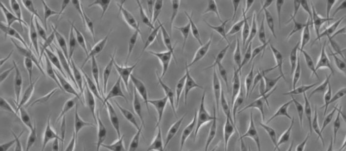
Figure 2. hiPSCs cultured on fibroblast-like feeder cells.
Overall, by modifying the extracellular environment, fibroblasts create a favorable environment for the growth of many different cell types.
Due to their ease of use, fibroblasts have been used for both primary cell cultures and for creating permanent, transformed cell lines. For example, the NIH3T3 cell line was derived from mouse embryonic fibroblasts transformed with the antigen T from SV40 virus; and has since been used in tens of thousands of publications2 .
Skin-derived fibroblasts are commonly used to produce induced pluripotent stem cells (iPSCs). iPSCs represent an important platform in cell culture studies because they can propagate indefinitely and give rise to any cell type in the body. Moreover, iPSCs allow the generation of the cell target of a particular disease while maintaining the genetic background of the patient of origin3 .
Consequently, fibroblasts can be used to produce iPSCs that represent powerful tools used for in vitro disease modelling.
As well as being a powerful tool for studying a limitless number of diseases in vitro through the generation of iPSCs, fibroblasts also offer a platform to explore the pathology of many fibroblast-related diseases directly.
Many fibroblast-related diseases are underscored by a process called fibrosis i.e. the overproduction of collagenous matrix, leading to the formation of scar tissue. Whilst this process is critical in sealing wounds, it can also be the basis of diseases such as systemic sclerosis (SSc), idiopathic pulmonary fibrosis (IPF), liver cirrhosis, kidney fibrosis, and cardiac fibrosis4 , when unregulated.
As well as their role in fibrosis, fibroblasts have been recognized as mediators of inflammation, disease-related inflammation and cancer progression. For example, fibroblasts maintain inflammation that leads to rheumatoid arthritis5 and supports tumor growth6 .
Due to their various roles in diseases, cultured fibroblasts can be used as model systems for many diseases and conditions. Additionally, cultured fibroblast cells have demonstrated utility in drug toxicology and screening. For example, cultured human skin fibroblasts have been used to investigate the cytotoxic effects of surfactants7 while lung fibroblast cell lines have been used to test the cytotoxicity of various antimycobacterial drugs8 .
Fibroblast growth factors (FGFs) represent a group of proteins produced by macrophages that signal fibroblasts to divide. Originally, FGF was thought to be important due to its effect on fibroblasts but they have since been recognized for their ability to stimulate many other types of cells. As such, FGFs have been extensively used in both 2D and 3D cell culture to promote cell proliferation, migration, differentiation, and angiogenesis9 .
Fibroblasts also play an important role in supporting one of the most influential developments in cell culture, which is the transition from 2D to 3D cultivation. Like 2D cell cultures, organoids can also benefit from the ability of fibroblasts to generate an optimal environment for cell growth. For instance, the addition of fibroblasts promoted the self-assembly of breast cancer cells into multicellular 3D structures. Compared to monocultures of breast cancer tissue cells that were absent of fibroblasts, the 3D cultures showed long-term viability and resistance to the chemotherapy drug paclitaxel10 .
Moreover, many organoids are generated from iPSCs derived from fibroblasts and require FGFs to help stimulate and direct growth.
It is becoming increasingly apparent that fibroblasts are advantageous cells for many 3D cell culture applications. As traditional 2D systems are increasingly replaced by 3D cell culture platforms , it is likely that fibroblasts will continue to retain their widespread use in cell culture studies.
3. They can be used to create permanent cell lines
Due to their ease of use, fibroblasts have been used for both primary cell cultures and for creating permanent, transformed cell lines. For example, the NIH3T3 cell line was derived from mouse embryonic fibroblasts transformed with the antigen T from SV40 virus; and has since been used in tens of thousands of publications2 .
4. Fibroblasts can be used to produce induced pluripotent stem cells (iPSCs)
Skin-derived fibroblasts are commonly used to produce induced pluripotent stem cells (iPSCs). iPSCs represent an important platform in cell culture studies because they can propagate indefinitely and give rise to any cell type in the body. Moreover, iPSCs allow the generation of the cell target of a particular disease while maintaining the genetic background of the patient of origin3 .
Consequently, fibroblasts can be used to produce iPSCs that represent powerful tools used for in vitro disease modelling.
5. Fibroblasts provide a model system for many diseases and conditions
As well as being a powerful tool for studying a limitless number of diseases in vitro through the generation of iPSCs, fibroblasts also offer a platform to explore the pathology of many fibroblast-related diseases directly.
Many fibroblast-related diseases are underscored by a process called fibrosis i.e. the overproduction of collagenous matrix, leading to the formation of scar tissue. Whilst this process is critical in sealing wounds, it can also be the basis of diseases such as systemic sclerosis (SSc), idiopathic pulmonary fibrosis (IPF), liver cirrhosis, kidney fibrosis, and cardiac fibrosis4 , when unregulated.
As well as their role in fibrosis, fibroblasts have been recognized as mediators of inflammation, disease-related inflammation and cancer progression. For example, fibroblasts maintain inflammation that leads to rheumatoid arthritis5 and supports tumor growth6 .
Due to their various roles in diseases, cultured fibroblasts can be used as model systems for many diseases and conditions. Additionally, cultured fibroblast cells have demonstrated utility in drug toxicology and screening. For example, cultured human skin fibroblasts have been used to investigate the cytotoxic effects of surfactants7 while lung fibroblast cell lines have been used to test the cytotoxicity of various antimycobacterial drugs8 .
Fibroblast Growth Factor and the Future of Fibroblasts
Fibroblast growth factors (FGFs) represent a group of proteins produced by macrophages that signal fibroblasts to divide. Originally, FGF was thought to be important due to its effect on fibroblasts but they have since been recognized for their ability to stimulate many other types of cells. As such, FGFs have been extensively used in both 2D and 3D cell culture to promote cell proliferation, migration, differentiation, and angiogenesis9 .
Fibroblasts also play an important role in supporting one of the most influential developments in cell culture, which is the transition from 2D to 3D cultivation. Like 2D cell cultures, organoids can also benefit from the ability of fibroblasts to generate an optimal environment for cell growth. For instance, the addition of fibroblasts promoted the self-assembly of breast cancer cells into multicellular 3D structures. Compared to monocultures of breast cancer tissue cells that were absent of fibroblasts, the 3D cultures showed long-term viability and resistance to the chemotherapy drug paclitaxel10 .
Moreover, many organoids are generated from iPSCs derived from fibroblasts and require FGFs to help stimulate and direct growth.
It is becoming increasingly apparent that fibroblasts are advantageous cells for many 3D cell culture applications. As traditional 2D systems are increasingly replaced by 3D cell culture platforms , it is likely that fibroblasts will continue to retain their widespread use in cell culture studies.
Read more
References
1. Caneparo, C. et al. Conditioned medium produced by fibroblasts cultured in low oxygen pressure allows the formation of highly structured capillary-like networks in fibrin gels. Sci. Rep. 10, (2020).
2. Leibiger, C. et al. First Molecular Cytogenetic High Resolution Characterization of the NIH 3T3 Cell Line by Murine Multicolor Banding. J. Histochem. Cytochem. 61, 306 (2013).
3. Fernandes, I. R. et al. Fibroblast sources: Where can we get them? Cytotechnology 68, 223 (2016).
4. Zhang, M. & Zhang, S. T Cells in Fibrosis and Fibrotic Diseases. Front. Immunol. 11, 1142 (2020).
5. Mizoguchi, F. et al. Functionally distinct disease-associated fibroblast subsets in rheumatoid arthritis. Nat. Commun. 2018 91 9, 1–11 (2018).
6. Winkler, J., Abisoye-Ogunniyan, A., Metcalf, K. J. & Werb, Z. Concepts of extracellular matrix remodelling in tumour progression and metastasis. Nat.Commun. 2020 111 11, 1–19 (2020).
7. JK, L., DB, K., JI, K. & PY, K. In vitro cytotoxicity tests on cultured human skin fibroblasts to predict skin irritation potential of surfactants. Toxicol. In Vitro 14, 345–349 (2000).
8. Takii, T. et al. Simple Fibroblast-Based Assay for Screening of New Antimicrobial Drugs against Mycobacterium tuberculosis. Antimicrob. Agents Chemother. 46, 2533 (2002).
9. Yun, Y.-R. et al. Fibroblast Growth Factors: Biology, Function, and Application for Tissue Regeneration. J. Tissue Eng. 2010, 1–18 (2010).
10. Jiang, T. et al. Bioprintable Alginate/Gelatin Hydrogel 3D In Vitro Model Systems Induce Cell Spheroid Formation. J. Vis. Exp. 2018, 57826 (2018).
Read more