MENÚ
AR | ARS

AR | ARS
-
- Todas las pipetas, dispensadores y manipuladores de líquidos automatizados
- Pipetas mecánicas
- Pipetas electrónicas
- Pipetas multicanal
- Pipetas y dispensadores de desplazamiento positivo
- Puntas de pipeta
- Dispensadores de botella
- Controladores de pipetas
- Accesorios para dispensadores y pipetas
- Pipeteo automatizado
- Consumibles para automatización
- Accesorios de automatización
- Servicios de manipulación de líquidos y pipetas
Sorry, we couldn't find anything on our website containing your search term.
Está a punto de abandonar este sitio.
Por favor, tenga en cuenta que su carro de la compra actual no ha sido guardado todavía y no podrá ser restablecido en el nuevo sitio ni cuando regrese. Si desea guardar su carro de la compra, inicie sesión en su cuenta.
Sorry, we couldn't find anything on our website containing your search term.
How to identify fungal contamination in your cell culture
Academia de laboratorio
- Biología celular
- Cultivo celular
- Contaminación
- Consumibles para cultivo celular
- Incubadores de CO2
- Ensayo
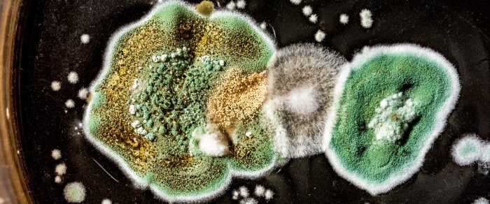
Macroscopic detection
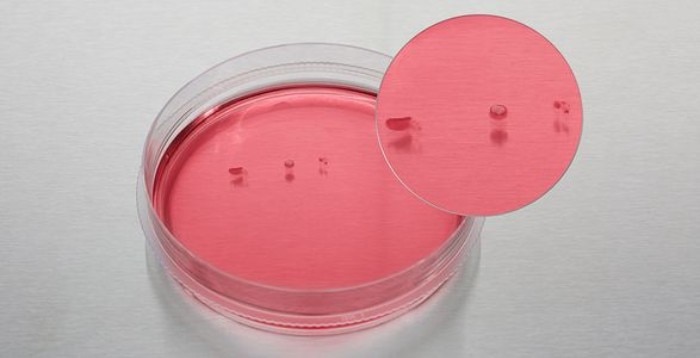
Fungi and mold can appear as small isolated colonies of grey, white or greenish color, floating at the surface of the medium. It is important to inspect the culture vessel with the naked eye and look for colonies which rest on the medium surface and will be missed during microscopic inspection. If the contamination is substantial, the medium will become turbid and cloudy, and spots on the vessel surface may appear. Sometimes fungal contaminations will cause a pH increase of the medium, resulting in phenol-red containing media to appear pink.Leer más
Fungal colonies floating on the medium surface.
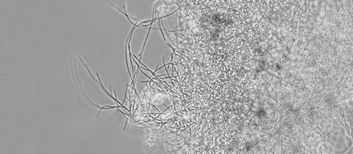
Microscopic detection
It is usually the fungal spores which get into a culture vessel and then begin to sprout and form the typical fiber like hyphae. Fungi growing on the bottom of the dish/flask are more easily detected in the microscope than those floating on the surface.Leer más
Related documents

White paper
PDF
PDF 0,90 MB
Related links
Leer más