MENU
BR | BRL

BR | BRL
-
- Todas as centrífugas
- Centrífugas de bancada
- Centrífugas de chão
- Centrífugas refrigeradas
- Microcentrífugas
- Centrífugas multifuncionais
- Centrífugas de alta velocidade
- Ultracentrífugas
- Concentrador
- Consumíveis de alta velocidade e de ultracentrifugação
- Tubos de centrifugação
- Placas de centrifugação
- Gestão de amostras e informações
-
- Todas as pipetas, dispensadores e equipamentos para manuseio de líquidos automatizados
- Pipetas mecânicas
- Pipetas eletrônicas
- Pipetas multicanal
- Pipetas de deslocamento positivo e dispensadores
- Ponteiras de pipeta
- Dispensadores de frascos
- Controladores de pipeta
- Acessórios para dispensadores e pipetas
- Pipetagem Automatizada
- Consumíveis de automação
- Acessórios de automação
- Serviços para pipetas e equipamentos para manuseio de líquidos
Sorry, we couldn't find anything on our website containing your search term.
Sorry, we couldn't find anything on our website containing your search term.
Into the Very Last Nerve Cell
Explore Life Science
- Lab Life
- Off the Bench
- Inspiring Science
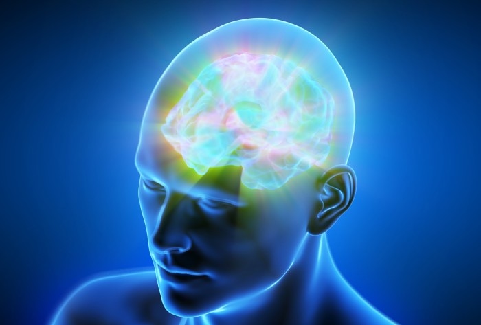
For the first time, we have high-definition 3D views of our brains. They deliver new insights into synaptic circuits.
One cubic millimeter equals a cube with sides each one millimeter long – that is the miniscule size of a tissue sample removed from a woman during epilepsy surgery. With the help of an electron microscope, the research team led by neuroscientist Jeff Lichtman from Harvard University was able to reveal astounding facts within that cube: 57,000 cells, including about 16,000 neurons, 23 centimeters of blood vessels and 150 million synapses. Even glial cells, responsible, among other things, for support and supply of neurons, could be detected.
In order to generate these views, the scientists cut the brain segment into fine layers, scanned these with an electron microscope, and reconstructed the single images of the layers into one 3D model. “The human brain is very complex. Currently, we know little about its cellular structure, particularly its so-called synaptic circuits,” say the researchers, and explain further that since these connections between nerve cells are often associated with neurological conditions, the model offers an important foundation for the understanding of those processes. Lichtman and his team hope that their model can make lasting contributions to a better grasp of the complex interactions in the human brain. In the meantime, laypeople can also see these brightly-colored structures for themselves: the 3D model is freely available online .
In order to generate these views, the scientists cut the brain segment into fine layers, scanned these with an electron microscope, and reconstructed the single images of the layers into one 3D model. “The human brain is very complex. Currently, we know little about its cellular structure, particularly its so-called synaptic circuits,” say the researchers, and explain further that since these connections between nerve cells are often associated with neurological conditions, the model offers an important foundation for the understanding of those processes. Lichtman and his team hope that their model can make lasting contributions to a better grasp of the complex interactions in the human brain. In the meantime, laypeople can also see these brightly-colored structures for themselves: the 3D model is freely available online .
Ler mais