MENU
FR | EUR
-
-
-
- Service pour pipette
- Services pour Mastercycler
- Services pour automates de pipetage
- Services pour bioprocédés
- Services pour congélateurs
- Services pour incubateurs
- Services de qualification
- Services pour agitateurs
- Service de contrôle de la température et de l’agitation
- Services pour centrifugeuse et rotors
-
-
- WHX Labs Dubai 2026
- analytica 2026
- Forum Labo 2026
- SLAS Europe 2026
- Forum Labo 2026 Bioprocess
- Bioprocessing Summit Europe 2026
- BioProcess International 2026
- Swiss Biotech Day 2025
- ISCT 2026
- Pichia 2026
- Future Labs Live 2026
- BioProScale Sympposium 2026
- ASGCT 2026
- Eppendorf Automation Forum 2026
- ESACT 2026
- ECB 2026
- WoTS 2026
- SIMB Annual Meeting 2026

-
-
-
- Service pour pipette
- Services pour Mastercycler
- Services pour automates de pipetage
- Services pour bioprocédés
- Services pour congélateurs
- Services pour incubateurs
- Services de qualification
- Services pour agitateurs
- Service de contrôle de la température et de l’agitation
- Services pour centrifugeuse et rotors
-
-
- WHX Labs Dubai 2026
- analytica 2026
- Forum Labo 2026
- SLAS Europe 2026
- Forum Labo 2026 Bioprocess
- Bioprocessing Summit Europe 2026
- BioProcess International 2026
- Swiss Biotech Day 2025
- ISCT 2026
- Pichia 2026
- Future Labs Live 2026
- BioProScale Sympposium 2026
- ASGCT 2026
- Eppendorf Automation Forum 2026
- ESACT 2026
- ECB 2026
- WoTS 2026
- SIMB Annual Meeting 2026
FR | EUR
-
- Pipettes, distributeurs et dispositifs de manipulation de liquides automatisée
- Pipettes mécaniques
- Pipettes électroniques
- Pipettes multicanaux
- Pipettes et distributeurs à déplacement positif
- Distributeurs sur flacon
- Auxiliaires de pipetage
- Accessoires pour pipettes et distributeurs
- Pipetage automatisé
- Consommables d’automatisation
- Accessoires d’automatisation
- Services pour pipettes et distributeurs
Sorry, we couldn't find anything on our website containing your search term.
Sorry, we couldn't find anything on our website containing your search term.
How to identify Mycoplasma contamination in your cell culture
Lab Academy
- Biologie cellulaire
- Culture cellulaire
- Contamination
- Consommables pour culture cellulaire
- Incubateurs à CO2
- Test
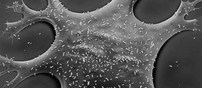
Macroscopic detection
Mycoplasma-positive cell cultures show no visible changes to the media.Microscopic detection
Mycoplasma are only about 0.1 - 0.3 µm in diameter, therefore detection via brightfield microscopy is not possible. This lack of visible signs of infection increases the risk of mycoplasma-positive cells remaining unnoticed.Experiments carried out with mycoplasma-infected cells may yield false, misleading and non-reproducible results. It is therefore crucial to test all cultures for mycoplasma on a regular basis. One simple method employs DNA staining; however, this method presents certain drawbacks. This table provides an overview of the advantages and disadvantages of different mycoplasma detection methods.
The method you will select may depend on one or more of the following:
- Access to the required equipment (for example thermocycler, fluorescence microscope, etc.).
- How many samples need to be tested at once.
- How urgently you need the results (the longer the testing procedure the higher the risk of spreading the contamination).
Lire la suite
Related documents

White paper
PDF
Overview
PDF 0,19 MB
Download
PDF 1,63 MB
Application Note
PDF
Related links
Lire la suite