MENU
IT | EUR
-
-
-
- Assistenza per pipette
- Assistenza per Mastercycler
- Assistenza per l'automazione
- Assistenza per bioprocessi
- Assistenza per ultracongelatori
- Assistenza per incubatori
- Servizi di qualificazione
- Assistenza per agitatori
- Assistenza per regolazione termica e miscelazione
- Assistenza per centrifughe e rotori
-
-
- WHX Labs Dubai 2026
- analytica 2026
- Forum Labo 2026
- SLAS Europe 2026
- Forum Labo 2026 Bioprocess
- Bioprocessing Summit Europe 2026
- BioProcess International 2026
- Swiss Biotech Day 2025
- ISCT 2026
- Pichia 2026
- Future Labs Live 2026
- BioProScale Sympposium 2026
- ASGCT 2026
- Eppendorf Automation Forum 2026
- ESACT 2026
- ECB 2026
- WoTS 2026
- SIMB Annual Meeting 2026

-
-
-
- Assistenza per pipette
- Assistenza per Mastercycler
- Assistenza per l'automazione
- Assistenza per bioprocessi
- Assistenza per ultracongelatori
- Assistenza per incubatori
- Servizi di qualificazione
- Assistenza per agitatori
- Assistenza per regolazione termica e miscelazione
- Assistenza per centrifughe e rotori
-
-
- WHX Labs Dubai 2026
- analytica 2026
- Forum Labo 2026
- SLAS Europe 2026
- Forum Labo 2026 Bioprocess
- Bioprocessing Summit Europe 2026
- BioProcess International 2026
- Swiss Biotech Day 2025
- ISCT 2026
- Pichia 2026
- Future Labs Live 2026
- BioProScale Sympposium 2026
- ASGCT 2026
- Eppendorf Automation Forum 2026
- ESACT 2026
- ECB 2026
- WoTS 2026
- SIMB Annual Meeting 2026
IT | EUR
-
- Tutte le pipette, i dispenser e i sistemi automatizzati per la manipolazione dei liquidi
- Pipette meccaniche
- Pipette elettroniche
- Pipette multicanale
- Pipette a spostamento positivo e dispenser
- Puntali per pipette
- Dispenser per flaconi
- Ausili di pipettaggio
- Accessori per dispenser e pipette
- Pipettaggio automatizzato
- Consumabili per l'automazione
- Accessori per l'automazione
- Servizi per sistemi di manipolazione dei liquidi e pipette
Sorry, we couldn't find anything on our website containing your search term.
Sorry, we couldn't find anything on our website containing your search term.

How to recognize bacterial contamination in your cell culture
Lab Academy
- Biologia cellulare
- Coltura cellulare
- Contaminazione
- Consumabili per colture cellulari
- Incubatori a CO2
- Saggio
This article was published first in "Inside Cell Culture" , the monthly newsletter for cell culture professionals. Find more interesting articles about CO2 incubators on our page "FAQs and material on CO2 incubators" .
Macroscopic detection
- Increased turbidity of culture medium, medium appears cloudy.
- If medium contains phenol red as pH indicator, a rapid color change to yellow indicates a sudden decrease in pH
Approfondisci
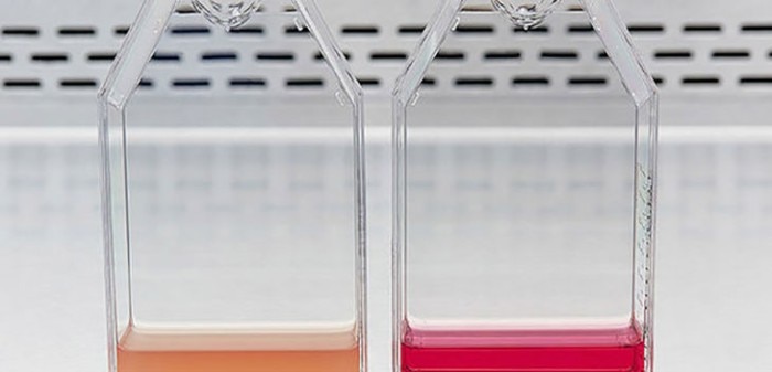
Culture media contaminated with bacteria (left) appear turbid and yellow whereas non-contaminated media (right) appear clear and red.
Microscopic detection
Bacteria are much smaller than eukaryotic cells. They appear as dark rod-like structures, spheres or spiral structures under the microscope, and they may exist as single cells, in pairs, chains, or clusters.Bacteria can be visualized using phase contrast and 100x - 400x magnification. Phase contrast facilitates detection, especially at low contamination levels.
Approfondisci

Common shapes of bacteria: rod (bacillus), spherical (coccus), and spiral (spirilla).
Moving bacteria in cell culture
Approfondisci
Videos not loading, because cookies have been rejected. Change your

Some bacteria show active movement. You are not sure whether what you see in your culture are cell debris or bacterial contamination? Try to focus on one of the potential contaminants and follow it with your eyes. Cell debris or any precipitates from the medium will only fidget but mostly stay in the same place. Some bacterial strains will actively move away from their current position.
Low levels of non-moving bacteria with round shapes are especially difficult to distinguish from any harmless particles fidgeting between the cells. In this case, only time can bring certainty: without antibiotics the number of particles will increase overnight if a contamination is present. If no increase occurs without antibiotics, most likely harmless debris or precipitates are at fault.
Low levels of non-moving bacteria with round shapes are especially difficult to distinguish from any harmless particles fidgeting between the cells. In this case, only time can bring certainty: without antibiotics the number of particles will increase overnight if a contamination is present. If no increase occurs without antibiotics, most likely harmless debris or precipitates are at fault.
Approfondisci

Related documents

White paper
PDF