메뉴
KR | KRW

KR | KRW
Sorry, we couldn't find anything on our website containing your search term.
How to detect a yeast contamination in your cell culture
실험실 아카데미
- 세포생물학
- Cell Culture
- 오염
- Cell Culture 소모품
- CO2 Incubator
- 논문
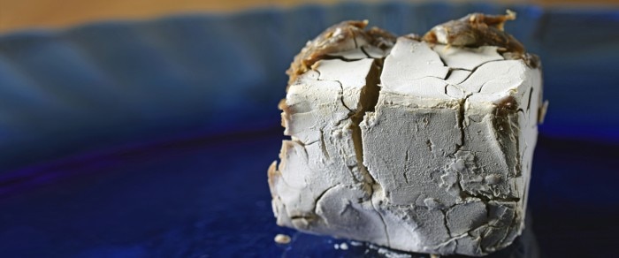
Macroscopic detection
- Increase in turbidity of the medium, medium becomes cloudy.
- Initial stages of yeast contamination are difficult to detect macroscopically, as the pH changes only slightly, initiating little or no color change in medium containing phenol red as pH indicator.
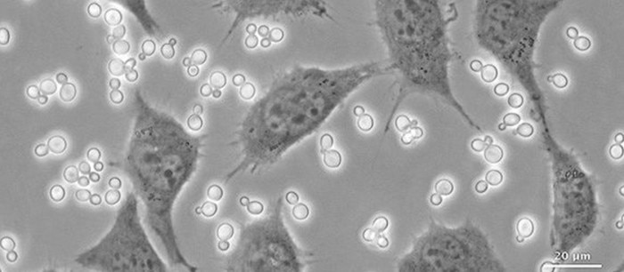
Microscopic detection
Yeasts can be visualized using phase contrast at 100x - 400x magnification. Phase contrast facilitates detection, especially at low contamination levels. Yeasts appear as ovoid bright particles between the cells. They can exist as single cells or in in the form of chains or branches.자세히 보기
Related documents

PDF 1.25 MB
Related links
자세히 보기